Testicular Malignant Mass || Ultrasound || Case 355
Testicular Malignant Mass || Ultrasound || Case 355
Visit for more: https://www.imagingstudy.com
Clinical Features:
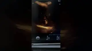
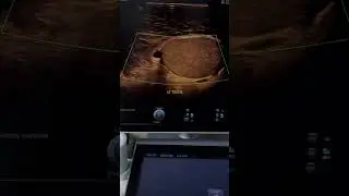
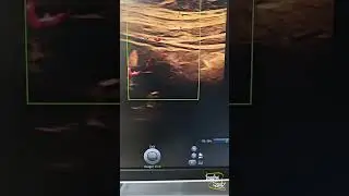
A 24-year-old male patient came with left testicular pain.
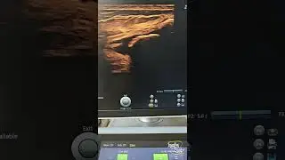
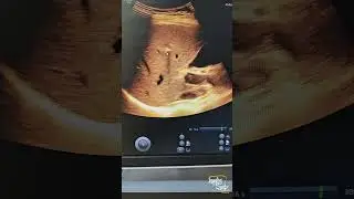
Ultrasound Features:
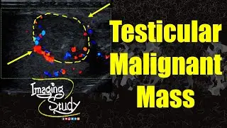
A solid, irregular, ill-defined heterogeneously hypoechoic mass is noted at the antero-inferior pole of the left testicular parenchyma with internal microcalcifications.
Predominantly peripheral vascularity is noted on color Doppler.
Remember:
Using different machine presets & transducers may help understand a lesion characteristic much better.
Thank you for watching.
Share with your friends.
Like & Subscribe for more videos.
Don't forget to put your valuable opinions in the Comment box below.
Check the playlist for Congenital Anomaly Lecture videos:
• Congenital Anomaly Lecture Series
Check the playlist for Imaging Study Lecture videos
• Imaging Study Lecture
Check the playlist for Imaging Study Ultrasound Case videos
• Ultrasound Cases
Follow us on
Facebook: / imagingstudychannel
Youtube: / imagingstudy
Instagram: / imagingstudy
Twitter: / imagingstudy
Telegram: https://t.me/ImagingStudy
Blogger: https://www.imagingstudy.com
#Ultrasound #Radiology #ImagingStudy #Ultrasoundcases #Medical #Imaging #Surgery #Mahin #Doctor #Medicine #reels #reel #short #youtube #facebook