Tongue Hemangioma || Ultrasound | Doppler || Case 351
Tongue Hemangioma || Ultrasound | Doppler || Case 351
Visit for more: https://www.imagingstudy.com
Clinical Features:
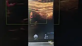
A 16-year-old male patient came with a gradually increasing bluish tongue swelling at the ventral aspect of the left lateral mid tongue.
Ultrasound Features:
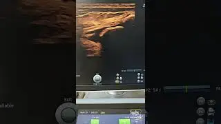
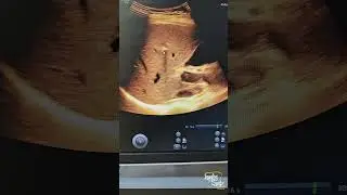
An oval hypoechoic area is noted at the ventral aspect of the left lateral mid tongue with internal prominent venous channels.
Color Doppler shows internal mild vascularity of both arterial & venous origins from adjacent tongue vessels.
Flow is seen within the venous channels on compression.
Surrounding tongue parenchyma appears uniform.
Remember:
Compressing a vascular malformation may demonstrate venous flow easily as they have got non-spontaneous flow commonly.
Thank you for watching.
Share with your friends.
Like & Subscribe for more videos.
Don't forget to put your valuable opinions in the Comment box below.
Check the playlist for Congenital Anomaly Lecture videos:
• Congenital Anomaly Lecture Series
Check the playlist for Imaging Study Lecture videos
• Imaging Study Lecture
Check the playlist for Imaging Study Ultrasound Case videos
• Ultrasound Cases
Follow us on
Facebook: / imagingstudychannel
Youtube: / imagingstudy
Instagram: / imagingstudy
Twitter: / imagingstudy
Telegram: https://t.me/ImagingStudy
Blogger: https://www.imagingstudy.com
#Ultrasound #Radiology #ImagingStudy #Ultrasoundcases #Medical #Imaging #Surgery #Mahin #Doctor #Medicine #reels #reel #short #youtube #facebook